The Burda Lab - Membrane Biology of Apicomplexan Parasites
Membranes are essential structural components of cells and play a central role in their function. They form barriers that separate the interior from the exterior in biological systems, and consist of a lipid bilayer embedded with proteins. Interactions between these lipids and proteins are important for membrane biogenesis, membrane organization, and the function of membrane proteins.
In our research group, we investigate the membrane biology of apicomplexan parasites during their replication in host cells. These single-cell parasites are of major medical and veterinary importance as they are responsible for a variety of diseases in humans and animals, including malaria, toxoplasmosis, and cryptosporidiosis. We are particularly interested in the mechanisms used by these parasites to build up, modify, and disrupt membranes to ensure their intracelluar multiplication. To understand these processes, we employ experimental genetics combined with high-resolution microscopy and biochemical approaches. This relatively unexplored field of research provides numerous new avenues for developing novel and urgently needed strategies to combat these intracellular pathogens.
Our current research focusses on the question how apicomplexan parasites acquire lipids from the host cell and how lipids are subsequently transported within the parasite. This includes not only the identification of the responsible molecular machinery of the parasites but also the characterization of potential host cell factors that might be hijacked by the parasites for this process.
Some examples of our previous research on the membrane biology of the malaria parasite:
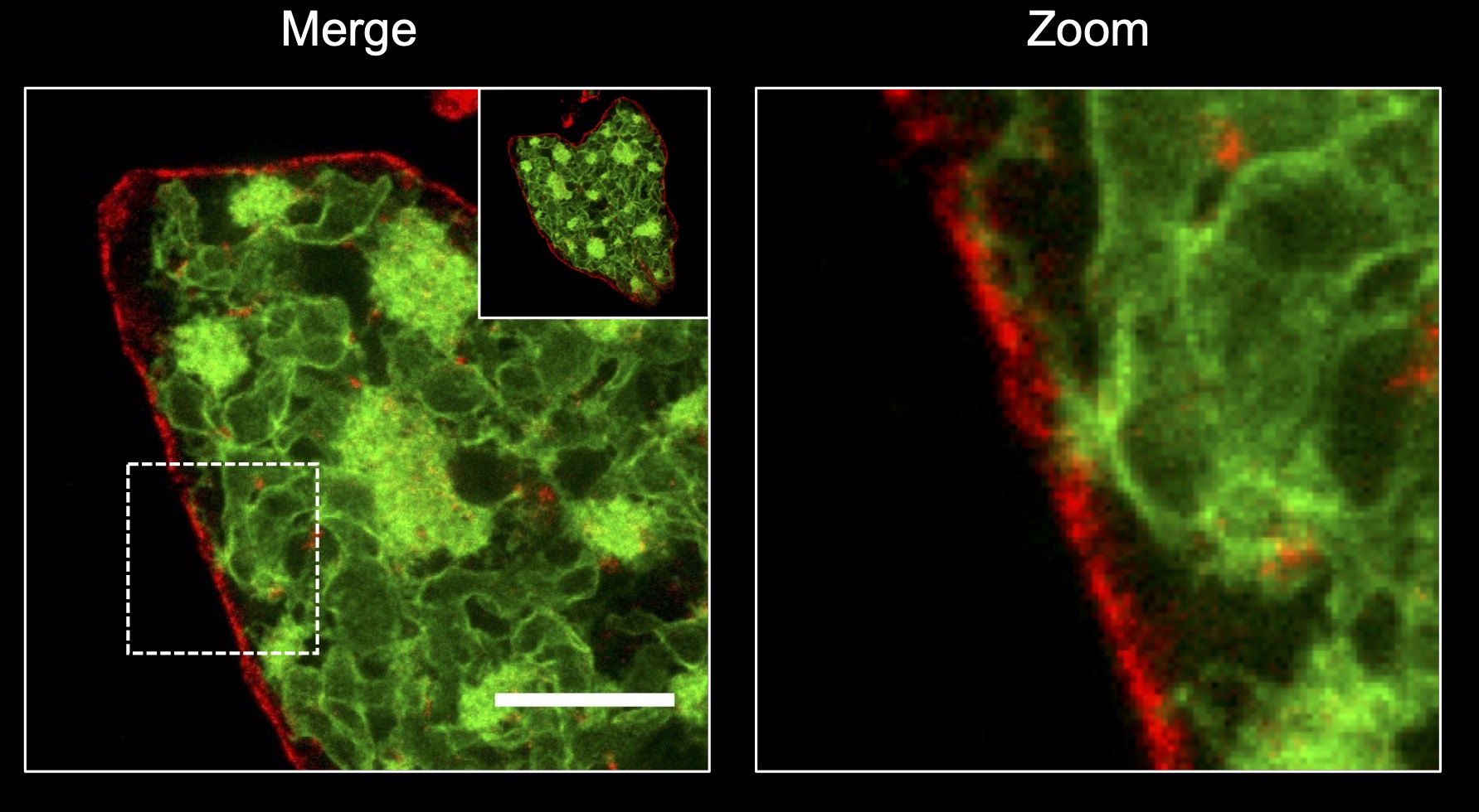
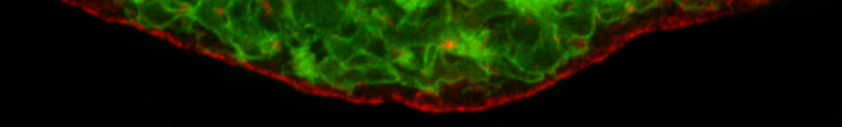
STED-based superresolution microscopy of a Plasmodium berghei liver stage stage parasites co-expressing a plasma membrane (red) and a endoplasmic reticulum (green) marker (Burda et al. 2017, Scientific Reports).
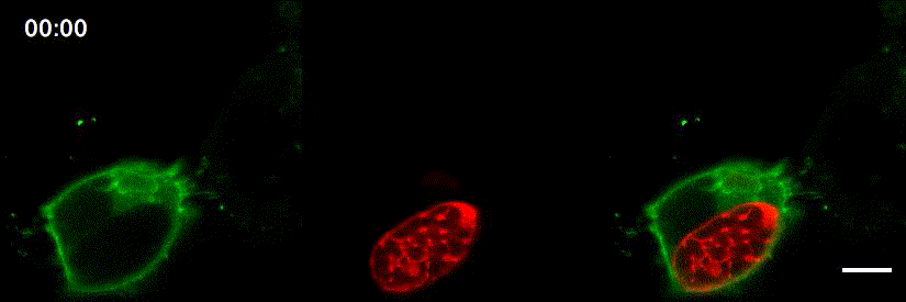
Confocal time-lapse microscopy of Plasmodium berghei liver stage egress. The parasitophorous vacuole membrane marker EXP1 is shown in red, while a host cell phosphatidylinositol 4,5-bisphosphate reporter is shown in green (Burda et al. 2017, mBio).
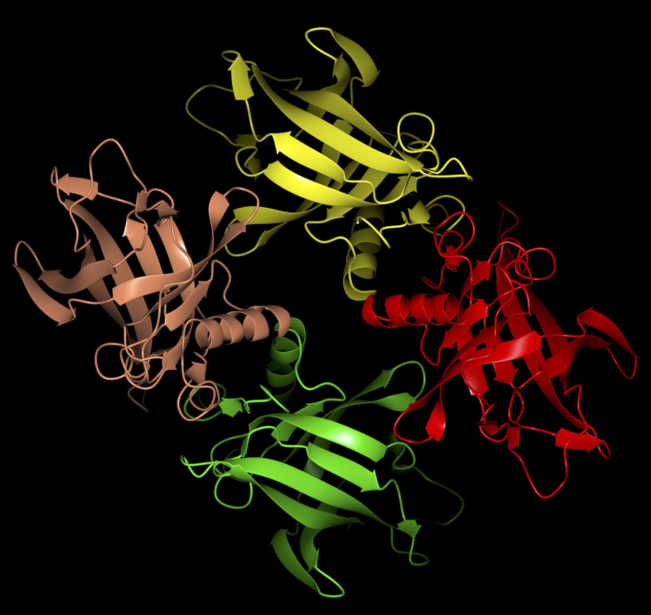
Crystal structure (PDB: 6TLB) of the Plasmodium falciparum lipocalin protein that plays an important function for replication of parasites within red blood cells (Burda et al. 2020, Cell Reports).
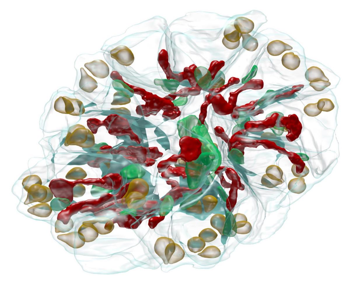
FIB-SEM-based 3D reconstruction of a Plasmodium falciparum schizont. Red, mitochondrion; green, apicoplast; light brown, rhoptries; light blue, parasite plasma membrane (Pietsch et al. 2023, mBio).
We acknowledge support by:


Membranes are essential structural components of cells and play a central role in their function. They form barriers that separate the interior from the exterior in biological systems, and consist of a lipid bilayer embedded with proteins. Interactions between these lipids and proteins are important for membrane biogenesis, membrane organization, and the function of membrane proteins.
In our research group, we investigate the membrane biology of apicomplexan parasites during their replication in host cells. These single-cell parasites are of major medical and veterinary importance as they are responsible for a variety of diseases in humans and animals, including malaria, toxoplasmosis, and cryptosporidiosis. We are particularly interested in the mechanisms used by these parasites to build up, modify, and disrupt membranes to ensure their intracelluar multiplication. To understand these processes, we employ experimental genetics combined with high-resolution microscopy and biochemical approaches. This relatively unexplored field of research provides numerous new avenues for developing novel and urgently needed strategies to combat these intracellular pathogens.
Our current research focusses on the question how apicomplexan parasites acquire lipids from the host cell and how lipids are subsequently transported within the parasite. This includes not only the identification of the responsible molecular machinery of the parasites but also the characterization of potential host cell factors that might be hijacked by the parasites for this process.
Some examples of our previous research on the membrane biology of the malaria parasite:
STED-based superresolution microscopy of a Plasmodium berghei liver stage stage parasites co-expressing a plasma membrane (red) and a endoplasmic reticulum (green) marker (Burda et al. 2017, Scientific Reports).
Confocal time-lapse microscopy of Plasmodium berghei liver stage egress. The parasitophorous vacuole membrane marker EXP1 is shown in red, while a host cell phosphatidylinositol 4,5-bisphosphate reporter is shown in green (Burda et al. 2017, mBio).
Crystal structure (PDB: 6TLB) of the Plasmodium falciparum lipocalin protein that plays an important function for replication of parasites within red blood cells (Burda et al. 2020, Cell Reports).
FIB-SEM-based 3D reconstruction of a Plasmodium falciparum schizont. Red, mitochondrion; green, apicoplast; light brown, rhoptries; light blue, parasite plasma membrane (Pietsch et al. 2023, mBio).
We acknowledge support by: